Imaging method reveals a “symphony of cellular activities” | MIT News

In just a solitary mobile, countless numbers of molecules, these types of as proteins, ions, and other signaling molecules, operate collectively to complete all sorts of functions — absorbing vitamins, storing recollections, and differentiating into unique tissues, among a lot of others.
Deciphering these molecules, and all of their interactions, is a monumental endeavor. More than the earlier 20 years, researchers have made fluorescent reporters they can use to go through out the dynamics of particular person molecules inside cells. Having said that, usually only one particular or two these indicators can be observed at a time, since a microscope can’t distinguish involving many fluorescent colors.
MIT scientists have now formulated a way to picture up to 5 distinct molecule varieties at a time, by measuring every single signal from random, unique destinations through a mobile. This method could allow for experts to discover considerably additional about the elaborate signaling networks that manage most mobile features, states Edward Boyden, the Y. Eva Tan Professor in Neurotechnology and a professor of organic engineering, media arts and sciences, and brain and cognitive sciences at MIT.
“There are 1000’s of molecules encoded by the genome, and they’re interacting in methods that we do not recognize. Only by seeing them at the exact same time can we recognize their interactions,” states Boyden, who is also a member of MIT’s McGovern Institute for Mind Exploration and Koch Institute for Integrative Most cancers Research.
In a new analyze, Boyden and his colleagues utilized this approach to determine two populations of neurons that respond to calcium indicators in distinctive ways, which could impact how they encode prolonged-term reminiscences, the researchers say.
Boyden is the senior creator of the examine, which seems these days in Mobile. The paper’s lead authors are MIT postdoc Changyang Linghu and graduate university student Shannon Johnson.
Fluorescent clusters
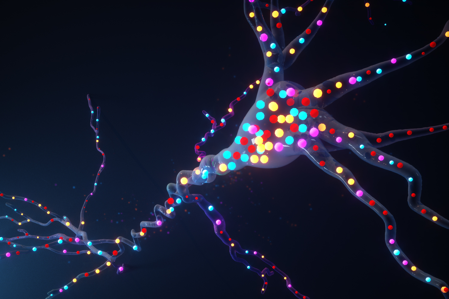
To make molecular exercise seen in a mobile, experts generally create reporters by fusing a protein that senses a goal molecule to a protein that glows. “This is related to how a smoke detector will feeling smoke and then flash a mild,” suggests Johnson, who is also a fellow in the Yang-Tan Centre for Molecular Therapeutics. The most typically used glowing protein is eco-friendly fluorescent protein (GFP), which is primarily based on a molecule at first identified in a fluorescent jellyfish.
“Typically a biologist can see just one or two shades at the identical time on a microscope, and a lot of of the reporters out there are environmentally friendly, simply because they are based on the environmentally friendly fluorescent protein,” Boyden says. “What has been lacking right until now is the ability to see a lot more than a couple of these alerts at at the time.”
“Just like listening to the audio of a single instrument from an orchestra is considerably from sufficient to absolutely take pleasure in a symphony,” Linghu states, “by enabling observations of various cellular indicators at the same time, our engineering will enable us have an understanding of the ‘symphony’ of cellular routines.”
To strengthen the variety of signals they could see, the scientists established out to determine signals by locale rather of by colour. They modified present reporters to bring about them to accumulate in clusters at distinctive areas within a cell. They did this by introducing two compact peptides to each reporter, which assisted the reporters sort distinct clusters inside cells.
“It’s like obtaining reporter X be tethered to a LEGO brick, and reporter Z tethered to a K’NEX piece — only LEGO bricks will snap to other LEGO bricks, triggering only reporter X to be clustered with more of reporter X,” Johnson states.
With this system, each and every cell finishes up with hundreds of clusters of fluorescent reporters. After measuring the activity of each and every cluster less than a microscope, based mostly on the shifting fluorescence, the scientists can identify which molecule was staying calculated in just about every cluster by preserving the cell and staining for peptide tags that are distinctive to just about every reporter. The peptide tags are invisible in the dwell cell, but they can be stained and viewed right after the live imaging is performed. This allows the scientists to distinguish indicators for distinctive molecules even although they may possibly all be fluorescing the same coloration in the stay cell.
Utilizing this solution, the scientists confirmed that they could see five various molecular indicators in a one cell. To demonstrate the probable usefulness of this tactic, they measured the things to do of 3 molecules in parallel — calcium, cyclic AMP, and protein kinase A (PKA). These molecules variety a signaling network that is included with numerous various mobile capabilities through the entire body. In neurons, it plays an important part in translating a shorter-phrase enter (from upstream neurons) into prolonged-time period changes these as strengthening the connections concerning neurons — a method that is vital for learning and forming new recollections.
Implementing this imaging system to pyramidal neurons in the hippocampus, the researchers identified two novel subpopulations with distinct calcium signaling dynamics. A person populace confirmed sluggish calcium responses. In the other population, neurons had a lot quicker calcium responses. The latter inhabitants experienced larger PKA responses. The researchers consider this heightened response may perhaps enable sustain long-long lasting improvements in the neurons.
Imaging signaling networks
The researchers now prepare to check out this solution in living animals so they can study how signaling network pursuits relate to habits, and also to expand it to other forms of cells, such as immune cells. This method could also be handy for evaluating signaling network patterns between cells from healthful and diseased tissue.
In this paper, the researchers showed they could history 5 unique molecular indicators at once, and by modifying their current tactic, they believe that they could get up to 16. With further do the job, that selection could arrive at into the hundreds, they say.
“That definitely may well enable crack open some of these hard questions about how the areas of a mobile perform with each other,” Boyden claims. “One may consider an era when we can watch anything heading on in a living cell, or at minimum the section associated with mastering, or with disorder, or with the remedy of a disease.”
The research was funded by the Pals of the McGovern Institute Fellowship the J. Douglas Tan Fellowship Lisa Yang the Yang-Tan Centre for Molecular Therapeutics John Doerr the Open up Philanthropy Venture the HHMI-Simons Faculty Scholars Software the Human Frontier Science System the U.S. Army Investigation Laboratory the MIT Media Lab the Picower Institute Innovation Fund the Countrywide Institutes of Health, like an NIH Director’s Pioneer Award and the National Science Basis.


